Home

dergi Etkileşim kendine hayranlık diffusion mri stroke fiil bir kere daha modül

MR-DWI In The Acute Stroke Diagnosis | STROKE MANUAL

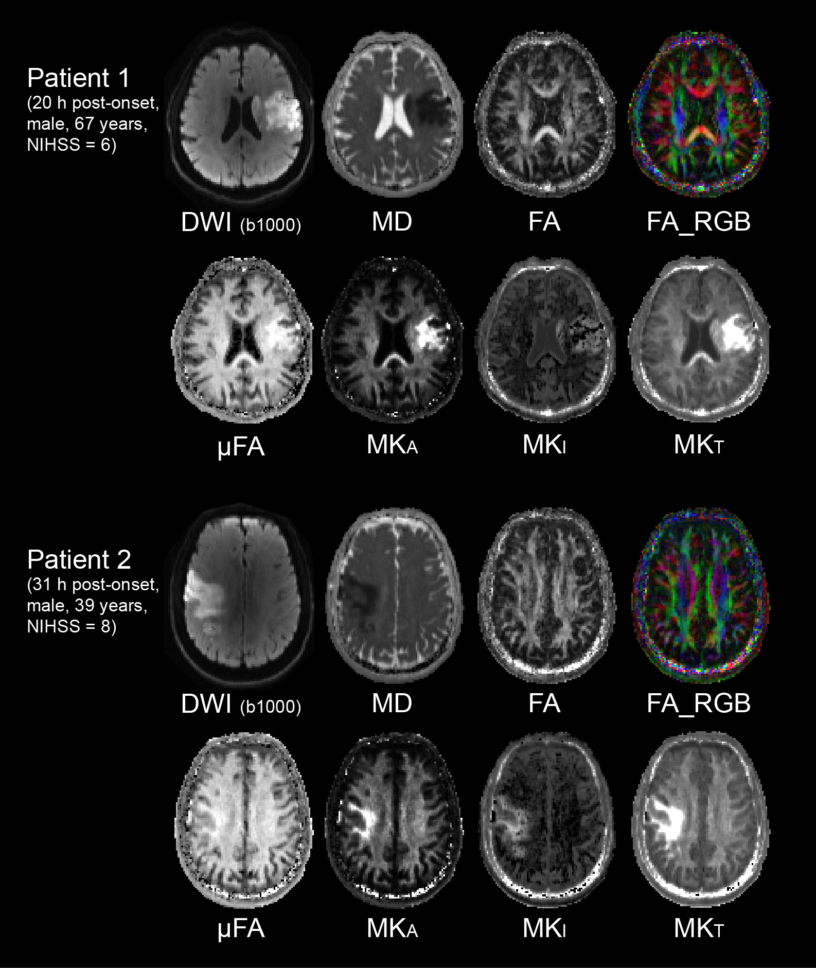
Identifying Severe Stroke Patients Likely to Benefit From Thrombectomy  Despite Delays of up to a Day | Scientific Reports
Identifying Severe Stroke Patients Likely to Benefit From Thrombectomy Despite Delays of up to a Day | Scientific Reports

Contribution of Diffusion-Weighted Imaging in Determination of Stroke  Etiology | American Journal of Neuroradiology
Contribution of Diffusion-Weighted Imaging in Determination of Stroke Etiology | American Journal of Neuroradiology

DWI - How Does Acute Infarct Cause Restricted Diffusion? - YouTube

Early Diffusion-Weighted Imaging Reversal After Endovascular Reperfusion Is  Typically Transient in Patients Imaged 3 to 6 Hours After Onset | Stroke
Early Diffusion-Weighted Imaging Reversal After Endovascular Reperfusion Is Typically Transient in Patients Imaged 3 to 6 Hours After Onset | Stroke

Diffusion-weighted MRI for evaluation of acute stroke | Neurology

PDF] Automatic Stroke Lesions Segmentation in Diffusion-Weighted MRI |  Semantic Scholar
PDF] Automatic Stroke Lesions Segmentation in Diffusion-Weighted MRI | Semantic Scholar

Clinical Applications of Diffusion MR Imaging for Acute Ischemic Stroke |  Radiology Key
Clinical Applications of Diffusion MR Imaging for Acute Ischemic Stroke | Radiology Key

Diffusion-weighted Imaging of Patients with Subacute Cerebral Ischemia:  Comparison with Conventional and Contrast-enhanced MR Imaging | American  Journal of Neuroradiology
Diffusion-weighted Imaging of Patients with Subacute Cerebral Ischemia: Comparison with Conventional and Contrast-enhanced MR Imaging | American Journal of Neuroradiology

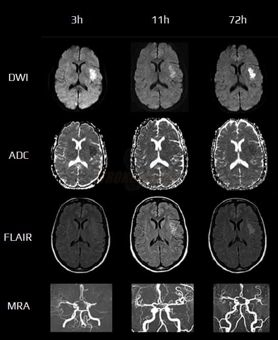
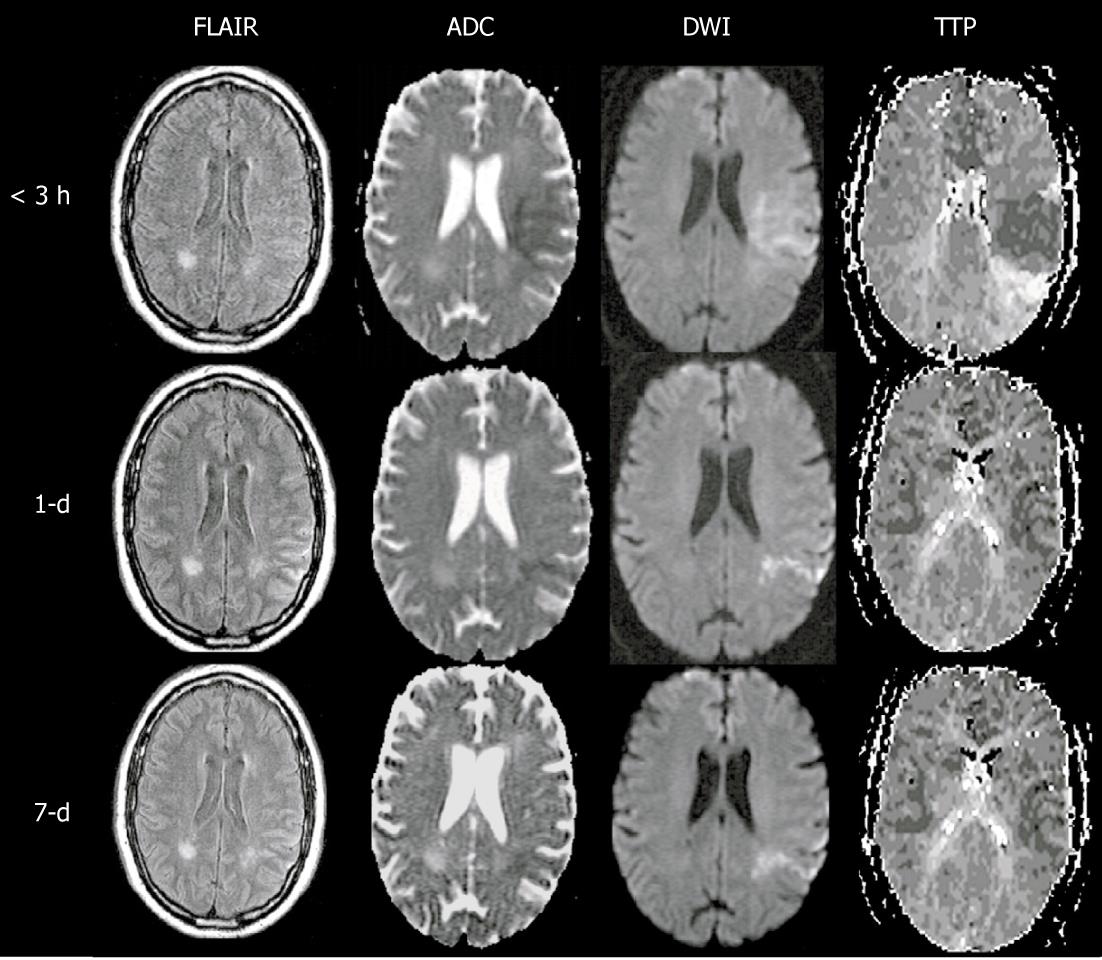
The diffusion weighted imaging (DWI) of 3 h stroke. (a) The... | Download  Scientific Diagram
The diffusion weighted imaging (DWI) of 3 h stroke. (a) The... | Download Scientific Diagram

Diffusion weighted imaging in acute ischemic stroke: A review of its  interpretation pitfalls and advanced diffusion imaging application -  ScienceDirect
Diffusion weighted imaging in acute ischemic stroke: A review of its interpretation pitfalls and advanced diffusion imaging application - ScienceDirect

Applications of diffusion–perfusion magnetic resonance imaging in acute  ischemic stroke | Neurology
Applications of diffusion–perfusion magnetic resonance imaging in acute ischemic stroke | Neurology

Diffusion weighted imaging in acute ischemic stroke: A review of its  interpretation pitfalls and advanced diffusion imaging application -  ScienceDirect
Diffusion weighted imaging in acute ischemic stroke: A review of its interpretation pitfalls and advanced diffusion imaging application - ScienceDirect

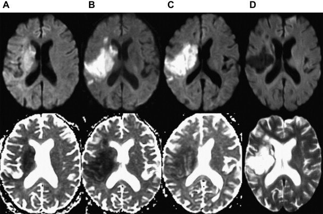
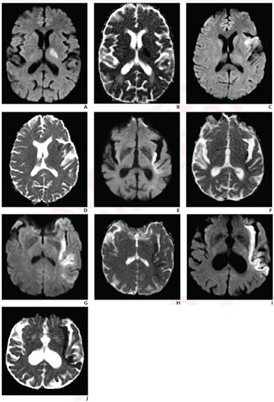
Figure 1 from Evolution of apparent diffusion coefficient, diffusion-weighted,  and T2-weighted signal intensity of acute stroke. | Semantic Scholar
Figure 1 from Evolution of apparent diffusion coefficient, diffusion-weighted, and T2-weighted signal intensity of acute stroke. | Semantic Scholar

Diffusion MRI of Cerebral Infarct | AccessMedicine Network

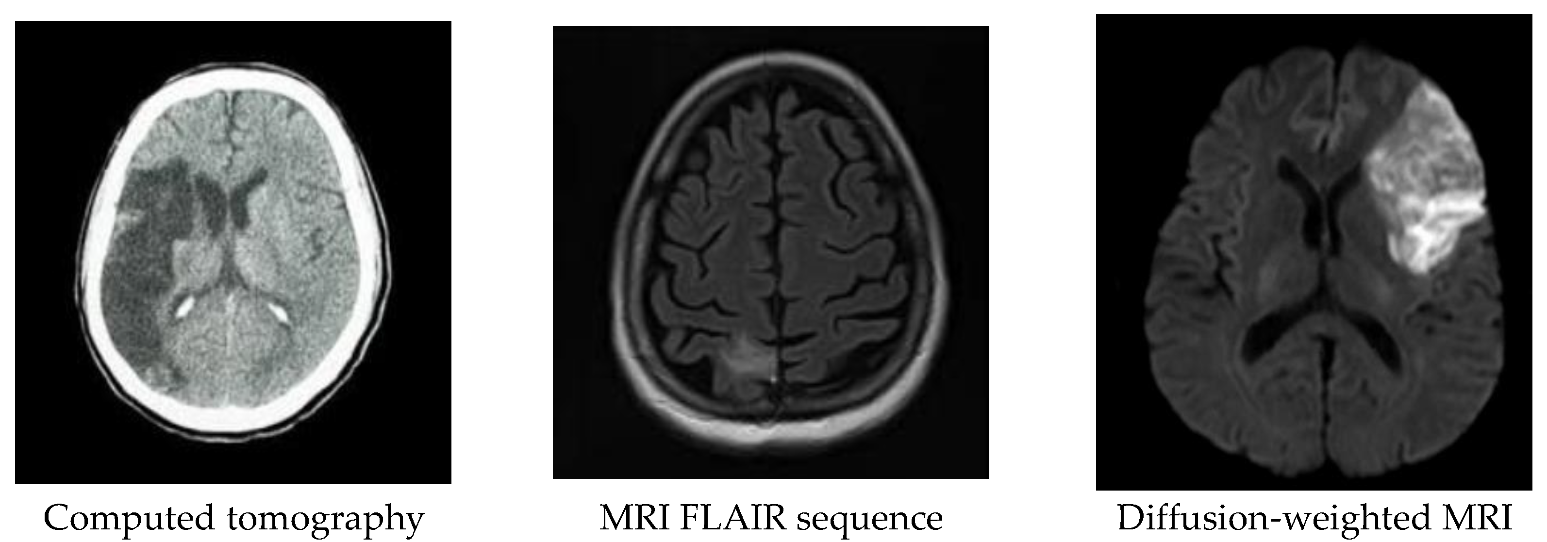
Diagnostics | Free Full-Text | Automatic Assessment of ASPECTS Using  Diffusion-Weighted Imaging in Acute Ischemic Stroke Using Recurrent  Residual Convolutional Neural Network
Diagnostics | Free Full-Text | Automatic Assessment of ASPECTS Using Diffusion-Weighted Imaging in Acute Ischemic Stroke Using Recurrent Residual Convolutional Neural Network

MR-DWI In The Acute Stroke Diagnosis | STROKE MANUAL

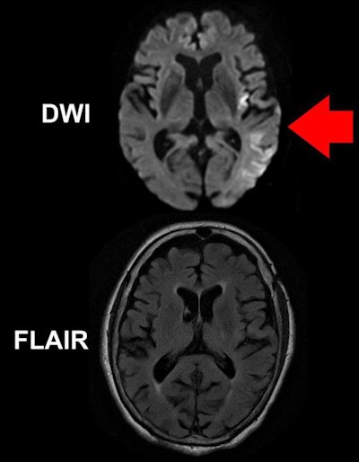
Stroke indicator found on MRI could help with treatment strategies |  AuntMinnie
Stroke indicator found on MRI could help with treatment strategies | AuntMinnie

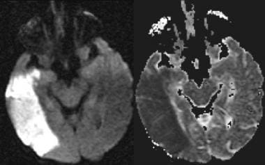
Diffusion-weighted MRI showing an acute large stroke in the right... |  Download Scientific Diagram
Diffusion-weighted MRI showing an acute large stroke in the right... | Download Scientific Diagram

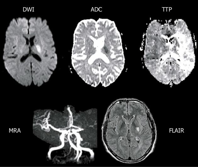
Comprehensive MRI assessment in acute stroke using DWI, PWI and MR... |  Download Scientific Diagram
Comprehensive MRI assessment in acute stroke using DWI, PWI and MR... | Download Scientific Diagram

Initial MRI after ischemic stroke A: Axial diffusion weighted imaging... |  Download Scientific Diagram
Initial MRI after ischemic stroke A: Axial diffusion weighted imaging... | Download Scientific Diagram

Magnetic resonance diffusion-perfusion mismatch in acute ischemic stroke:  An update
Magnetic resonance diffusion-perfusion mismatch in acute ischemic stroke: An update

ISMRM2021-000526_Fig2.jpg

Sensitivity of Diffusion- and Perfusion-Weighted Imaging for Diagnosing  Acute Ischemic Stroke Is 97.5% | Stroke
Sensitivity of Diffusion- and Perfusion-Weighted Imaging for Diagnosing Acute Ischemic Stroke Is 97.5% | Stroke

Application of Diffusion – And Perfusion – Weighted Imaging in Acute  Ischemic Stroke | IntechOpen
Application of Diffusion – And Perfusion – Weighted Imaging in Acute Ischemic Stroke | IntechOpen

Diagnostics | Free Full-Text | Application of Machine Learning Techniques  for Characterization of Ischemic Stroke with MRI Images: A Review
Diagnostics | Free Full-Text | Application of Machine Learning Techniques for Characterization of Ischemic Stroke with MRI Images: A Review

Clinical applications of diffusion-weighted sequence in brain imaging:  beyond stroke | Neuroradiology
Clinical applications of diffusion-weighted sequence in brain imaging: beyond stroke | Neuroradiology

Magnetic resonance diffusion-perfusion mismatch in acute ischemic stroke:  An update
Magnetic resonance diffusion-perfusion mismatch in acute ischemic stroke: An update